
When you notice an unusual spot, bump, or sore inside your child’s mouth, it’s natural to feel concerned. These oral changes can appear suddenly and range from completely harmless to requiring professional attention. Pediatric dental cleanings play an important role in identifying oral lesions early, allowing our team to monitor changes and recommend appropriate care when needed.
At Pine Tree Pediatric Dentistry, Dr. Tesha Waggoner understands that oral lesions in children can be a source of worry for parents. With compassionate care and thorough evaluations, we help families navigate common pediatric oral conditions while providing guidance and treatment to maintain healthy smiles.
Understanding Oral Lesions in Children
Oral lesions are changes in the color, size, or structure of the normal oral anatomy that can appear anywhere inside the mouth. Research from the American Academy of Pediatric Dentistry indicates that the most frequently observed oral mucosal lesions in children and adolescents include aphthous ulcerations, herpes simplex virus infections, trauma-associated lesions, benign migratory glossitis (geographic tongue), and candidiasis. While these conditions may cause concern for parents, many are benign and resolve without intervention.
Children’s oral lesions can present as white patches, red areas, ulcers, bumps, or other changes to the normal pink, smooth appearance of oral tissues. Some lesions cause pain and discomfort, while others remain completely asymptomatic. Understanding the different types helps parents know when to seek professional evaluation.
Viral and Fungal Oral Lesions
Several infectious conditions can create oral lesions in children. These include viral infections, such as herpes simplex virus, and fungal infections, such as oral candidiasis. Both types respond well to appropriate treatment when identified early.
Herpes Simplex Virus Infection
Primary herpetic gingivostomatitis is the most common symptomatic presentation of an initial herpes simplex virus infection in children. This condition presents as red, bleeding gums and clusters of small blisters throughout the mouth. Children may experience fever, difficulty eating, and swollen lymph nodes. The condition is self-limiting, with symptoms typically resolving within two weeks, though topical treatments can provide relief for discomfort.
Oral Candidiasis
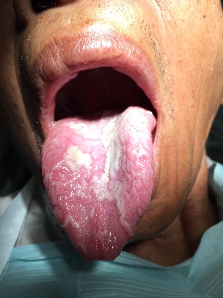
Oral candidiasis is a relatively common fungal infection that appears as white patches or plaques covering the mouth tissues. When removed, these patches leave an underlying surface that appears red or irritated. This condition may affect infants, children taking certain medications, or those with compromised immune systems. Healthy children may not require treatment if the condition is asymptomatic, though antifungal medications are available when needed.
What Are Recurrent Aphthous Ulcerations?
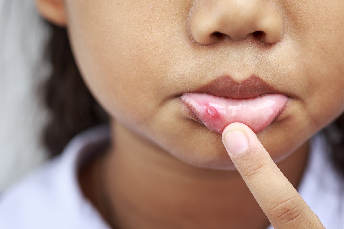
Aphthous ulcers, commonly called canker sores, occur in 20 to 30 percent of children. These painful lesions appear primarily on nonkeratinized tissues, such as the insides of the cheeks, lips, and the underside of the tongue. The ulcers typically have a yellowish-white center surrounded by a red border.
Minor aphthous ulcerations are the most common form, measuring 3 to 10 millimeters in diameter. Between one and five ulcers often present during a single outbreak and heal within 7 to 10 days without scarring. Treatment focuses on pain relief through topical anesthetics, with corticosteroids serving as first-line treatment for more severe cases. Parents should encourage children to avoid spicy or acidic foods that may irritate the ulcers during healing.
Common Reactive and Developmental Lesions
Several oral lesions develop as reactions to trauma or as part of normal oral development. A mucocele is a common lesion in children resulting from damage to a minor salivary gland, often due to lip biting or trauma. These appear as blue, red, or translucent swellings, most commonly on the lower lip. Small mucoceles usually resolve spontaneously, while larger ones may require surgical removal.
Traumatic injuries can also lead to oral lesions. An irritation fibroma develops as a response to chronic irritation of the mouth tissues and presents as a firm, pink nodule typically found on the cheeks, lips, or tongue. Removing the source of irritation and surgically removing the fibroma prevents recurrence.
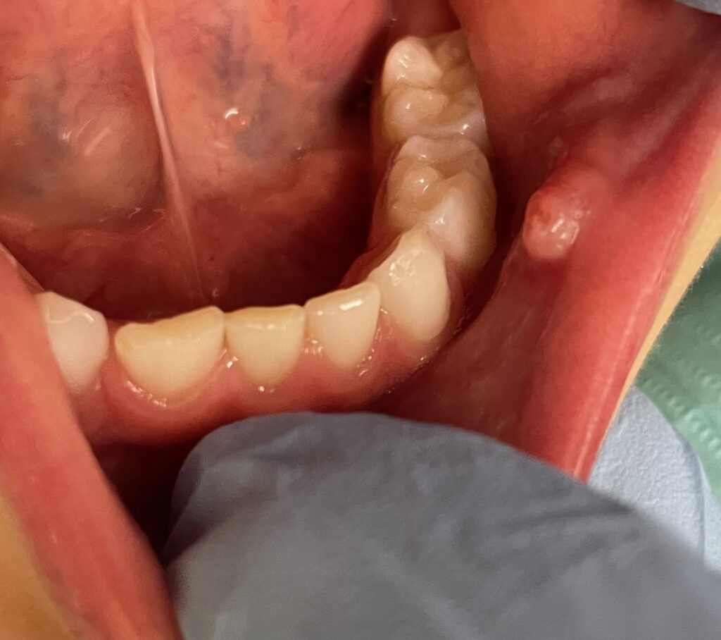
Eruption cysts sometimes appear as a tooth prepares to emerge through the gum tissue. These soft tissue cysts range in color from normal to blue-black, depending on the amount of fluid present. They most commonly occur with the eruption of the first permanent molars and front teeth. Treatment is rarely necessary, as the tooth naturally erupts through the cyst.
When to Seek Professional Evaluation
Parents should schedule an evaluation when an oral lesion persists for more than two weeks without improvement, causes significant pain or difficulty eating, appears to be growing, or is accompanied by fever or other systemic symptoms. Our experienced team can perform a thorough examination to establish a diagnosis and recommend appropriate treatment.
During routine dental visits, we carefully examine all oral tissues to identify any unusual findings early. This proactive approach allows us to monitor changes over time and intervene when necessary. For children with anxiety about dental examinations, our office offers helpful accommodations, including our therapy dog Meyers, weighted blankets, and the option to watch a movie during appointments.
Trust Pine Tree Pediatric Dentistry for Comprehensive Oral Health Care
At Pine Tree Pediatric Dentistry, we’re committed to helping families understand and manage common oral conditions in children. Our compassionate approach combines thorough clinical evaluation with patient education, empowering parents to make informed decisions about their child’s oral health.
Whether your child needs routine preventive care or evaluation of an oral lesion, learn more about our practice or contact our office to schedule an appointment today.